| 中心波长 | 780 nm | 1560 nm |
| 平均输出功率 | > 140 mW | > 350 mW |
| 脉宽 | < 100 fs | < 100 fs |
| 重复率 | 标准值 80 MHz* | |
| 光束发散角 | < 1 mrad | < 2 mrad |
| 光束尺寸 (1/e²) | Ø 1.2 mm (典型值) | Ø 3.5 mm (典型值) |


 基于LabVIEW的图形用户界面 软件控制功能:内置功率PC控制系统,通过网络页面轻松交流,可直接访问电动控制装置(例如不同的脉冲压缩设置),LabVIEW驱动模板便于系统集成
 FemtoFiber pro 控制单元配备单键开关,钥匙开关,连锁开关,12英寸机架外壳包含接口,泵浦二极管的驱动电子元件以及电源。
| M40: 重复率 40 MHz (而非标准版本的 80 MHz) | |
| Mxx: 客户定制重复率,例如 68 MHz, 77 MHz, … | |
| AMP: 多光束系统 | 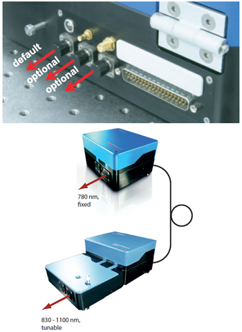 |
| VAR: 可调重复率 | 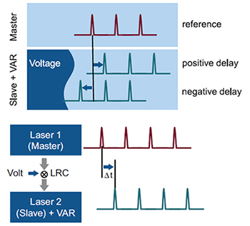 |
| LRC: 激光重复率控制 | 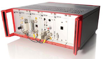 |
| 1ps: 脉冲长度拓展至皮秒量级 | 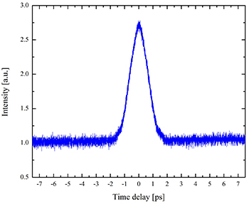 |
| 附加种子端口,为多线输出装置做准备 | |
| 根据需求可提供1560 nm及780 nm两波长间分光比固定或可变设置,而非简单的机械开关。 | |
适用于 FemtoFiber pro NIR 以及 TNIR 系统
脉冲长度典型值:1 ps (NIR/780 nm), 0.5 - 1ps (TNIR/850-1100 nm)
方形脉冲
其余产品规格可另行调整
可选用替换晶体
锁相环路电子装置,以将激光脉冲序列与外部参考信号或激光参照系统相同步
信号抖动均方根 < 200 fs
紧凑型电子机架,配备电源
USB 接口及控制软件
系统可根据根据振荡单元调节,实现重复频率调制
通过高速控制压电传感器调节谐振腔长度,谐振频率> 1 kHz
利用慢速控制电动载物台,可实现典型可调范围200kHz(标称重复率± 100 kHz范围)
拓展系统配备放大器,但并无振荡器(因此称为“AMP”,即amplifier)
主振荡器系统的外部种子源通过FC/APC光纤接口输入
主振荡器可根据情况增加额外的种子源端口
多光束系统,可支持多达4台系统集成
FemtoFiber pro 系列中所有系统搭配可选
流行的多光束系统:
主振荡器型号AMP 1 型号应用目标NIRTNIR AMPCARS 激光源NIRUCP AMP宽带 CARS 光源IRSCIR AMPOPCPA 种子系统TVISTVIS AMP泵浦探针光谱
振荡器设计支持40MHz重复率
部分规格可能有所变动(详情请联系我们)
与VAR选项不兼容
双光子聚合
多光子显微镜
二次谐波(SHG)显微镜
太赫兹产生
Jr, C.H.C., et al., High-speed coherent Raman fingerprint imaging of biological tissues. Nat. Photonics 8, 627–634 (2014)
Galli, R. et al. Vibrational Spectroscopic Imaging and Multiphoton Microscopy of Spinal Cord Injury. Anal. Chem. 84, 8707–8714 (2012)
Galli, R. et al. Non-linear optical microscopy of kidney tumours. J. Biophotonics n/a–n/a (2013)
Krauss, G. et al. Compact coherent anti-Stokes Raman scattering microscope based on a picosecond two-color Er:fiber laser system. Opt. Lett. 34, 2847–2849 (2009)
Galli, R. et al. Intrinsic Indicator of Photodamage during Label-Free Multiphoton Microscopy of Cells and Tissues. PLoS ONE 9, e110295 (2014)
Paar, M. et al. Remodeling of Lipid Droplets during Lipolysis and Growth in Adipocytes. J. Biol. Chem. 287, 11164–11173 (2012)
Galli, R. et al. Effects of tissue fixation on coherent anti-Stokes Raman scattering images of brain. J. Biomed. Opt. 19, 071402–071402 (2013)
Galli, R. et al. CARS and non-linear microscopy imaging of brain tumors. 87970E–87970E (2013).
Chun, W., et al. Design and demonstration of multimodal optical scanning microscopy for confocal and two-photon imaging. Rev. Sci. Instrum. 84, 013701 (2013)
Jeong, H.-J., et al. Spectrally resolved fluorescence lifetime imaging microscope using tunable bandpass filters. Rev. Sci. Instrum. 83, 093705–093705–5 (2012)